Get Healthy!

- Posted July 28, 2023
New Ultrasound Patch Spots Tiny Breast Abnormalities in Early Trial
Scientists have developed a wearable ultrasound patch that might eventually allow women to monitor themselves for early signs of breast cancer in the comfort of their home.
The achievement, reported July 28 in the journal Science Advances, is the latest in a broader research effort to make wearable ultrasound a reality.
The hope is to one day use such portable technology to help diagnose and monitor a range of diseases and injuries -- in a way that's more accessible and cheaper than using traditional scanners housed at medical facilities.
The U.S. National Institutes of Health, which is funding some of that research, says that wearable ultrasound has the potential to "revolutionize health care."
Right now, breast ultrasound is used to help detect cancer in some women. If a screening mammogram picks up a suspicious finding, for example, ultrasound may be done to see whether it's a tumor or a cyst. Ultrasound can also be used, in addition to screening mammography, when a woman has particularly dense breast tissue (which makes it harder for radiologists to see a tumor on a mammogram).
That, however, requires women to go to a health care facility, and the ultrasound test itself is "operator-dependent," explained senior researcher Canan Dagdeviren, an associate professor at the Massachusetts Institute of Technology (MIT) .
With traditional breast ultrasound, a health care provider applies a gel to a handheld wand -- called a transducer -- then moves it over the skin on and around the breast. So, the quality of one ultrasound to another varies, in part, based on the operator's experience and expertise.
In theory, a wearable ultrasound device could be both more convenient and more reliable.
But the breast presents a particular design challenge: curves. Other wearable ultrasound devices under development have typically been small -- even the size of a postage stamp -- and designed to be used once, for a matter of days.
That approach is believed to hold promise for imaging the heart, arteries, lungs or other organs and tissue as people go about their day.
But Dagdeviren is interested in a different use: Can wearable ultrasound, used repeatedly over the long term, help catch breast cancer in its earliest stages -- particularly in women at high risk?
Breast cancer screening is already routine for women starting in their 40s, but it's only done periodically. That, Dagdeviren said, leaves the problem of "interval cancers" -- breast cancers that develop after a normal screening mammogram.
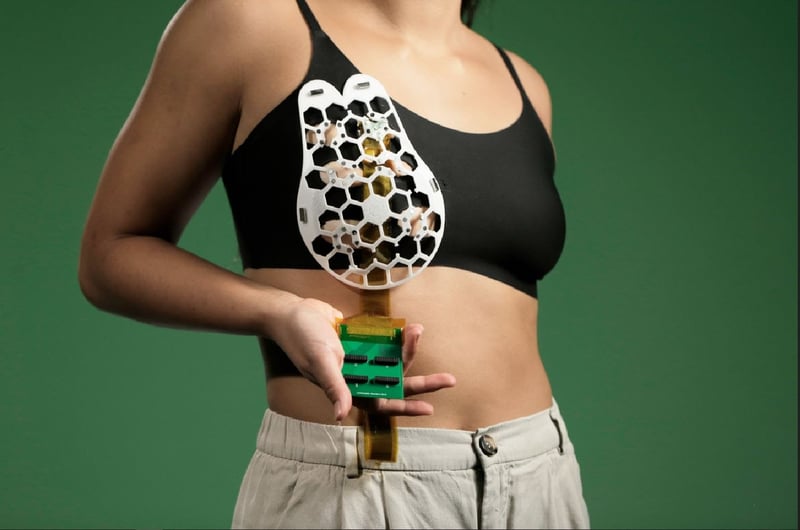
"So we thought, why don't we create a malleable, flexible ultrasound device that you can wear over your bra?" Dagdeviren said.
Her team has done just that. It's what Dagdeviren describes as a honeycomb-like patch, with 15 hexagonal sections. That makes it flexible enough to conform to the breast, over a bra, and also gives it structure: Ultrasound images are captured by a tiny tracker that moves around the patch, via a set pathway along the hexagons. The tracker can rotate 360 degrees, providing images at multiple angles.
The researchers put the patch to an early test with the help of a 71-year-old woman who had a history of cysts in her breast tissue. They found that the device could detect cysts as small as 3 millimeters -- hinting at its potential to pick up early tumors.
"This is a very impressive study," said Sheng Xu, a researcher at the University of California, San Diego, whose lab has been developing wearable ultrasound for a number of years.
"The tracker in the bra could help standardize the ultrasound imaging procedure and minimize the operator-dependency, which plagues conventional ultrasound," Xu said.
There's much work left to be done, however. Dagdeviren said her team will need to study at least 1,000 women to win approval for the patch from the U.S. Food and Drug Administration.
And for the initial testing, the patch was attached by a cable to a standard computer, to display the ultrasound images. That obviously won't work for home use, Dagdeviren said. So, the researchers want to pair the patch with a smartphone-like device; the data would be sent via cloud to a health care provider.
Xu and his team recently reported that they've developed a fully wireless ultrasound patch -- one that can monitor the carotid artery in the neck, measuring blood pressure, artery stiffness and how well the heart is pumping blood to the brain.
Dagdeviren said the breast ultrasound patch could potentially be used by women at high risk of breast cancer, due to factors like strong family history. She also envisions it providing an option for women in lower-income countries, where access to conventional medical imaging is limited.
"We're working on making this technology affordable," Dagdeviren said. "This is very important to me."
She and a colleague on the work are the inventors on a patent related to the ultrasound patch.
More information
The U.S. National Institutes of Health has more on wearable ultrasound.
SOURCES: Canan Dagdeviren, PhD, associate professor, media arts and sciences, Massachusetts Institute of Technology, Cambridge, Mass.; Sheng Xu, PhD, associate professor, nano-engineering, Jacobs School of Engineering, University of California San Diego; Science Advances, July 28, 2023, online


